A stained specimen of bacteria to study morphology sets the stage for this enthralling narrative, offering readers a glimpse into a story that is rich in detail and brimming with originality from the outset. This comprehensive guide delves into the fascinating world of bacterial morphology, providing a detailed exploration of the techniques, applications, and significance of this field.
Through meticulous preparation, staining, and microscopic examination, scientists unlock the secrets of bacterial structure, gaining invaluable insights into their physiology, pathogenicity, and ecological roles. Join us on this scientific expedition as we uncover the hidden beauty and profound implications of bacterial morphology.
Specimen Preparation

Preparing a stained specimen of bacteria for morphological study involves several steps to ensure optimal visualization and preservation of bacterial morphology. The first step is to collect the bacterial sample, which can be obtained from various sources such as clinical specimens, environmental samples, or pure cultures.
The sample is then subjected to a fixation process, typically using chemical fixatives like formaldehyde or glutaraldehyde, to preserve the bacterial morphology and prevent degradation.
Staining Technique
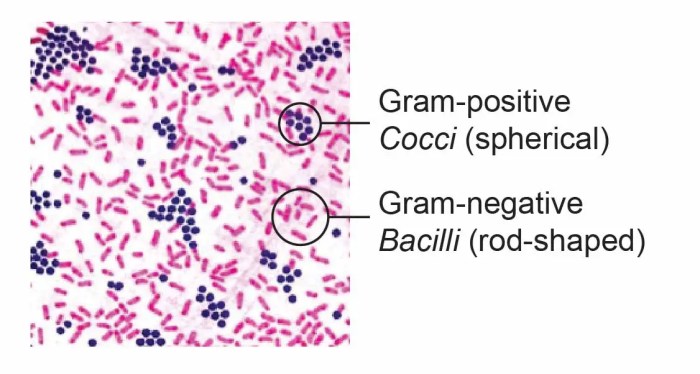
Once the sample is fixed, staining techniques are employed to enhance the visualization of bacterial morphology under a microscope. The choice of staining technique depends on the specific characteristics of the bacteria being studied. Gram staining is a widely used technique that differentiates bacteria into two groups based on their cell wall structure.
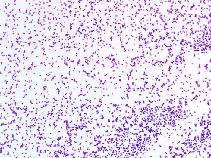
Gram-positive bacteria retain the crystal violet stain, appearing purple, while Gram-negative bacteria lose the stain and are counterstained with safranin, appearing pink or red. Other staining techniques include acid-fast staining, which is used to identify bacteria with a waxy cell wall, and fluorescent staining, which utilizes fluorescent dyes to label specific bacterial components.
During specimen preparation, specific considerations and precautions must be taken to ensure accurate and reliable results. These include maintaining proper sample collection and handling techniques to prevent contamination or alteration of the bacterial morphology. Additionally, the staining procedure must be carefully followed, and the appropriate controls must be included to ensure the validity of the results.
Morphological Analysis

Morphological analysis of bacteria involves examining the physical characteristics of bacteria, including their shape, size, arrangement, and the presence of specific structures. Microscopy techniques allow researchers to visualize these morphological features and gain insights into bacterial identification, classification, and physiology.
Morphological Characteristics
The shape of bacteria can vary greatly, including spherical (cocci), rod-shaped (bacilli), spiral (spirilla), and filamentous (spirochetes). The size of bacteria is typically measured in micrometers (µm) and can range from a few hundred nanometers to several micrometers. The arrangement of bacteria can also provide valuable information, such as pairs (diplococci), chains (streptococci), or clusters (staphylococci).
Staining Techniques
Staining techniques play a crucial role in enhancing the visualization of morphological characteristics. Gram staining, for example, not only differentiates bacteria based on their cell wall structure but also reveals the presence of endospores, which are dormant structures found in some bacterial species.
Acid-fast staining highlights bacteria with a waxy cell wall, which is a characteristic of Mycobacterium species. Fluorescent staining allows for the visualization of specific bacterial components, such as flagella or pili, which are essential for bacterial motility and attachment.
Identification and Classification
Morphological features are essential for identifying and classifying bacteria. By observing the shape, size, arrangement, and presence of specific structures, researchers can narrow down the possible bacterial species. For example, the presence of endospores is a defining characteristic of the genus Bacillus, while the spiral shape is a characteristic of the genus Spirillum.
Morphological analysis, combined with other techniques such as biochemical tests and molecular analysis, enables accurate identification and classification of bacteria.
Microscopy Techniques
Microscopy techniques are essential tools for studying bacterial morphology. Different microscopy techniques offer varying levels of resolution and magnification, allowing researchers to visualize different aspects of bacterial structure.
Light Microscopy
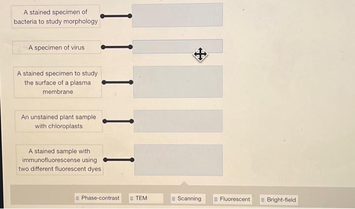
Light microscopy is a widely used technique that employs visible light to illuminate the specimen. Bright-field microscopy is a basic light microscopy technique that provides a clear view of bacterial morphology. Phase-contrast microscopy enhances the contrast of transparent specimens, making it useful for observing unstained bacteria.
Dark-field microscopy utilizes oblique lighting to create a dark background, highlighting the edges of bacterial cells.
Electron Microscopy
Electron microscopy offers much higher resolution than light microscopy, allowing for the visualization of ultrastructural details. Transmission electron microscopy (TEM) uses a beam of electrons to penetrate the specimen, providing detailed images of internal structures. Scanning electron microscopy (SEM) scans the surface of the specimen with a beam of electrons, creating three-dimensional images of the bacterial surface.
Selection of Technique
The choice of microscopy technique depends on the specific research objectives. Light microscopy is suitable for basic morphological studies, while electron microscopy is necessary for examining ultrastructural details. Researchers must consider the resolution and magnification required, as well as the potential impact of sample preparation on bacterial morphology.
Image Analysis
Image analysis techniques play a crucial role in quantifying and analyzing morphological features of bacteria. These techniques allow researchers to extract precise measurements and data from microscopic images.
Software and Tools
Various software and tools are available for image analysis, including ImageJ, CellProfiler, and MicrobeJ. These software packages provide a range of image processing and analysis capabilities, such as image enhancement, segmentation, and feature extraction.
Quantification of Features
Image analysis enables the quantification of morphological features, such as cell size, shape, and arrangement. By applying appropriate algorithms, researchers can measure the length, width, and area of individual bacterial cells. They can also determine the circularity, eccentricity, and other shape descriptors.
Applications in Research, A stained specimen of bacteria to study morphology
Image analysis is widely used in bacterial morphology research. It allows researchers to compare the morphological characteristics of different bacterial strains, study the effects of environmental conditions on bacterial morphology, and investigate the relationship between morphology and bacterial function.
Applications of Morphological Analysis: A Stained Specimen Of Bacteria To Study Morphology

Morphological analysis of bacteria has broad applications in various fields, including microbiology, medicine, and environmental science.
Bacterial Physiology
Morphological characteristics can provide insights into bacterial physiology. For example, the presence of flagella or pili indicates the ability of bacteria to move, while the presence of endospores indicates their ability to survive harsh conditions.
Pathogenicity
Morphological features can be associated with bacterial pathogenicity. Certain shapes and arrangements are more commonly associated with pathogenic bacteria, and the presence of specific structures, such as capsules or fimbriae, can enhance the virulence of bacteria.
Ecology
Morphological analysis is used to study the diversity and distribution of bacteria in different environments. The shape, size, and arrangement of bacteria can vary depending on the environmental conditions, and morphological analysis can provide insights into the ecological roles of bacteria.
Answers to Common Questions
What is the significance of studying bacterial morphology?
Bacterial morphology plays a crucial role in understanding bacterial physiology, pathogenicity, and ecological interactions. Morphological characteristics can provide insights into a bacterium’s ability to adhere to surfaces, invade host cells, and respond to environmental cues.
How do staining techniques enhance the visualization of bacterial morphology?
Staining techniques employ dyes that selectively bind to specific components of bacterial cells, such as the cell wall, cytoplasm, or flagella. This differential staining allows for clear visualization and differentiation of different bacterial structures under a microscope.
What are the different microscopy techniques used to study bacterial morphology?
Various microscopy techniques are employed to study bacterial morphology, including bright-field microscopy, dark-field microscopy, phase-contrast microscopy, and electron microscopy. Each technique offers unique advantages and limitations, depending on the specific research objectives and the level of detail required.